
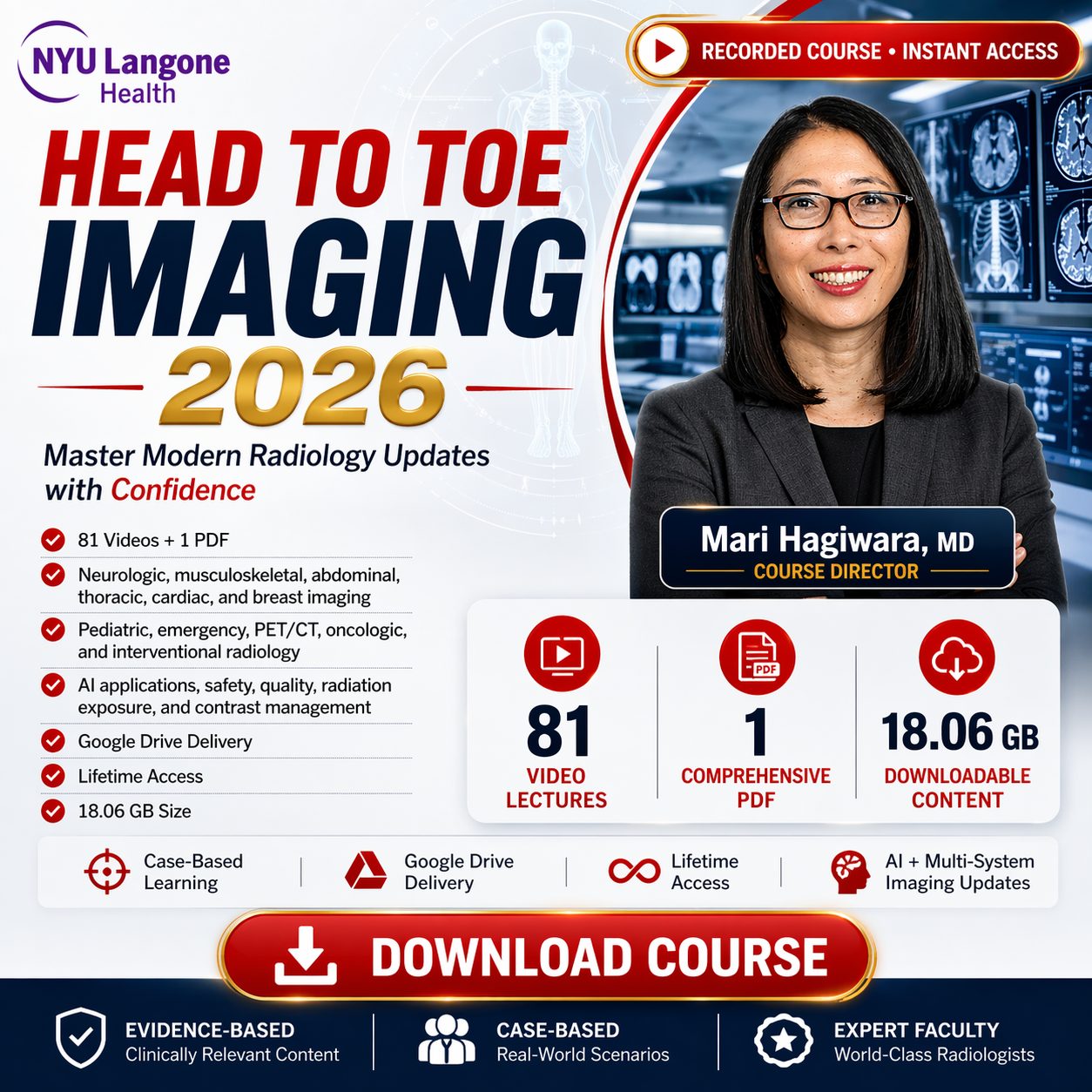

Head to Toe Imaging
Explore the Latest in Radiology Imaging
Stay at the forefront of radiology with NYU Langone's Head to Toe Imaging, a comprehensive program built for today’s evolving imaging landscape. This case-based online video program delivers state-of-the-art learning that translates directly to clinical practice.
What You'll Gain
- The latest updates in neurologic, musculoskeletal, abdominal, thoracic, cardiac, and breast imaging.
- Integrated coverage of pediatric, emergency, PET/CT, oncologic imaging, and interventional radiology.
- Guidance on safety, quality, radiation exposure, and contrast reaction management.
- Insight into emerging AI applications and standardized imaging strategies.
Strengthen diagnostic confidence and deliver high-quality, evidence-based patient care.
Topics & Speakers
Cardiothoracic Imaging
- Pulmonary Artery CTA and Multi-Energy Imaging Jane P. Ko, MD
- The Challenging Acute Aorta Smita Patel, MBBS, MRCP, FRCR
- Coronary Arteries – Beyond Atherosclerosis Smita Patel, MBBS, MRCP, FRCR
- Cardiac Pathology Identified in Non-Cardiac Non-Gated Studies Panagiota Christia, MD
- Q&A Session Faculty
- Cardiac Valvular Disease Kana Fujikura, MD, MPH
- Challenging Cardiac CT Cases Jill E. Jacobs, MD
- Evaluation of Cardiac Function Leon Axel, MD, PhD
- Q&A Session Faculty
- Lung Cancer Screening Dexter P. Mendoza, MD
- Imaging of Lung Cancer Charlotte Charbel, MD
- Primary and Metastatic Lung Tumor Interventions Nathan K. Mickinac, MD
- Q&A Session Faculty
- Chest MR William H. Moore, MD
- Esophageal Emergencies Gene Y. Berkovich, MD
- ICU Plain Film Jadranka Stojanovska, MD
- Q&A Session Faculty
Breast Imaging
- Method of Detection of Breast Cancer, the New Biomarker Jean M. Seely, MD
- Update on Management of BBD with a Focus on High-Risk Lesions Samantha L. Heller, MD, PhD
- AI in Breast Imaging Jean M. Seely, MD
- Tumor Board for the Radiologist Beatriu Reig, MD, MPH
- Updates on ACR Appropriateness Criteria for Screening Alana Lewin, MD
- Q&A Session Faculty
- CEM and MRI – Strengths, Limitations, and Synergies Michelle Lee, MD
- Auditing Breast MRI Jean M. Seely, MD
- Breast MRI Safety Julia Po, MD
- Latest and Greatest in Breast Imaging You Can Use Linda Moy, MD
- Q&A Session Faculty
Abdominal Imaging
- De-Mystifying the Mesentery Myles Taffel, MD
- Ovarian Imaging – Current Approaches and Advances Kira Melamud, MD, MHPE
- Abdominal Ultrasound – Pearls and Pitfalls Daniel T. Parrott, MD
- Easy to Fly By Incidental Critical Findings in Body Imaging Jay Karajgikar, MD
- Best Body Club Cases of The Year Sarah R. Beier, MD
- Q&A Session Faculty
- Pancreatic Cyst Imaging and Evolving Guidelines Chenchan Huang, MD
- Pancreatic Neuroendocrine Tumor – Updates in Imaging and Management Linda Chu, MD
- DOTA-peptide PET – Essentials and Challenges Marius E. Mayerhoefer, MD, PhD
- AI in Body Imaging – Where Are We Now Linda Chu, MD
- Q&A Session Faculty
- Liver Imaging – LI-RADS and Beyond Krishna P. Shanbhogue, MD
- Liver Tumor Ablation – A Review of Standard and Emerging Technologies Mikhail Silk, MD
- Rad-Path Discordance in Prostate MRI – Impact of Image Quality Angela Tong, MD
- Q&A Session Faculty
- Renal Masses – Updates and the Role of KI-RADS Nicole M. Hindman, MD
- Pitfalls of Bowel Interpretation on Routine Abdominal and Pelvic CT Douglas S. Katz, MD
- Pediatric Emergency Abdominal Imaging Naomi A. Strubel, MD
- Q&A Session Faculty
Neuroradiology
- Stroke Code – Challenging Cases and Lessons Learned Evan G. Stein, MD, PhD
- Brain Tumor Basics in the Molecular Era Roshni R. Patel, DO
- Susceptibility Weighted Imaging – My Favorite Sequence After DWI Rajan Jain, MD
- Spinal CSF Leak – What the Radiologist Needs to Know Peter G. Kranz, MD
- Q&A Session Faculty
- Case-Based Neuroanatomy Andrew C. McClelland, MD, PhD
- Not Futile Anymore – Imaging of Alzheimer's Disease in the Era of Disease Modifying Therapy James R. Loftus, MD
- Imaging of the Post-Operative Spine (Made Easy) Peter G. Kranz, MD
- Q&A Session Faculty
- Fractures of Child Abuse – What the Radiologist Needs to Know Shailee V. Lala, MD
- Abusive Head Trauma – Brain to Bones – Imaging Insights in Suspected Child Abuse Kevin Hsu, MD
- Head and Neck Emergencies Mari Hagiwara, MD
- Dotatate PET in Neuroradiology – Meningiomas and Paragangliomas Kent P. Friedman, MD
- Q&A Session Faculty
- Upper Aerodigestive Tract Made Easy Gopi K. Nayak, MD
- Navigating Complex Facial Trauma Matthew S. Breen, MD
- Neuro Jeopardy – Test Your Knowledge Evan Lieberman, MD
- Q&A Session Faculty
Musculoskeletal Imaging
- Classification Systems for Soft Tissue Tumors – How to Incorporate Into Practice Hillary Garner, MD
- Knee Chondral Joint-Preserving Surgical Options: MRI Assessment Erin Alaia, MD
- Imaging of Musculoskeletal Interventions – An update Using a Case-based Approach Christopher J. Burke, MD
- Muscle Injuries Iman Khodarahmi, MD, PhD
- Q&A Session Faculty
- High Water Content Soft Tissue Masses Hillary Garner, MD
- Spinal Trauma – Key Issues in the Emergent Setting David Y. Kim, MD
- Musculoskeletal Manifestations of Cancer and Treatment-Related Conditions Anton Becker, MD, PhD
- Q&A Session Faculty
- Vascular Anomalies – Diagnostic Imaging and Interventional Approaches Frederic J. Bertino, MD & Alexander El-Ali, MD
- Imaging of Arthroplasties Meghan Jardon Zikaras, MD
- Q&A Session Faculty
- Get Hip to It – Pelvis and Hip Cases Dana Lin, MD
- Hand and Wrist Challenging Cases Sophie L. Leung, MD
- Foot and Ankle Challenging Cases Alexandra Napolitano, MD
- Q&A Session Faculty
Program Faculty
Course Director

Mari Hagiwara, MD
Professor, Department of Radiology
NYU Grossman School of Medicine
New York, NY
Guest Speakers

Linda Chu, MD
Associate Professor
Johns Hopkins University, Lutherville, MD

Hillary Garner, MD, FACR
Professor of Radiology
Mayo Clinic, Neptune Beach, FL

Peter George Kranz, MD
Associate Professor with Tenure
Duke University, Durham, NC

Smita Patel, MBBS, MRCP, FRCR
Professor
University of Michigan, Saline, MI

Jean M. Seely, MD
Professor of Radiology
University of Ottawa, Ottawa, Ontario
NYU Grossman School of Medicine Speakers
Assistant Professor
Professor
Assistant Professor
Assistant Professor
Assistant Professor
Assistant Professor
Assistant Professor
Associate Professor
Assistant Professor
Assistant Professor
Assistant Professor
Assistant Professor
Associate Professor
Professor
Professor
Assistant Professor
Associate Professor
Professor
Professor
Assistant Professor
Associate Professor
Professor
Associate Professor
Associate Professor
Professor
Associate Professor
Assistant Professor
Assistant Professor
Associate Professor
Assistant Professor
Assistant Professor
Assistant Professor
Professor
Assistant Professor
Associate Professor
Associate Professor
Assistant Professor
Professor
Professor
Assistant Professor
Assistant Professor
Assistant Professor
Assistant Professor
Assistant Professor
Assistant Professor
Professor
Assistant Professor
Professor
Associate Professor
Associate Professor
Professor
Associate Professor
Program Details
Target Audience
The program is designed for clinical radiologists in either general or specialized practice, and is appropriate for radiologists in training. Familiarity with the fundamentals of CT, MR, PET, and US technology and applications, as well as basic anatomy, is assumed.
Educational Objectives
After participating in this activity, participants should be able to:
- Highlight key differences in Photo-counting CT image acquisition and image reconstruction compared with conventional CT. To summarize early evidence for the clinical benefit of PCCT for high-spatial resolution diagnostic tasks in thoracic imaging, such as assessment of airway and parenchymal diseases. Describe the benefits of high-pitch and multienergy scanning, including the ability for radiation dose reduction, depending on the diagnostic task. Evaluate the applications of PCCT for thoracic imaging in children. Describe the limitations and trade-offs of PCCT. Provide an overview of the best ways to incorporate PCCT into clinical practice including prioritizing access to patients and diagnostic tasks most likely to benefit from PCCT technology.
- Evaluate the MRI features of the most common chondral repair tissue techniques, including microfracture, autologous chondrocyte implantation, osteochondral allograft, and osteochondral autograft transfer. To recognize important complications after chondral repair surgery. To understand the features of the MOCART scoring system to assess the quality of chondral repair tissue.
- Describe the radiographic characteristics of ARIA that occur with anti-amyloid therapy in patients with Alzheimerʼs disease.
- Recognize the complexity of breast cancer screening and the need to optimize risk-based testing. To incorporate evidence-based best practices in providing supplemental breast cancer screening in light of the recent federal legislation. To describe the rationale and understand how to document method of detection (MOD) in breast cancer patients.
- Describe the imaging characteristics and differential diagnoses of common pancreatic cystic lesions, accounting for key demographic information. Evaluate cyst features associated with increased risk of malignancy based on current guidelines. Consider integrating abbreviated protocol into pancreatic cyst surveillance.
What Your Peers Are Saying
"Exceptional coverage of the latest in breast imaging. The AI applications segment was particularly eye-opening and directly applicable to my daily practice."
- Dr. Sarah J., Radiologist
"High-yield, case-based learning. Exactly what I needed for my practice. The thoracic imaging updates were stellar and the case examples were complex but clear."
- Dr. Mark D., Clinical Radiologist
"A phenomenal review of head to toe imaging. The faculty from NYU Langone are truly top-notch and the platform is incredibly easy to use."
- Dr. Emily R., Attending Physician
Frequently Asked Questions
Can I watch the videos on my mobile device?
Yes! Because the course is delivered via Google Drive, you can easily stream or download the videos on your smartphone or tablet using the Google Drive app or your mobile web browser.
How long will I have access to the materials?
You will receive lifetime access to all course materials, meaning you can review the content and use it as an ongoing reference for your clinical practice indefinitely.
Can I view the content offline?
Yes, you have the option to securely download the video files and the accompanying PDF syllabus for times when you don't have an active internet connection.
Ready to Upgrade Your Practice?
Get instant access to hours of state-of-the-art radiology education and comprehensive case-based learning materials from NYU Langone Health.
Reviews
There are no reviews yet.